Blog

Sciatica nerve pain – affecting the deep six muscles, the hip rotators.

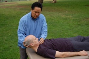
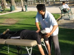
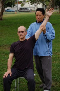
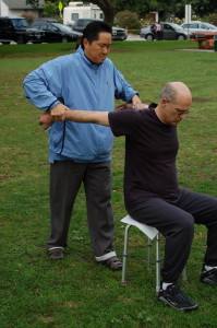
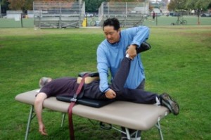
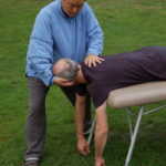
Sciatica nerve pain involves compression on the sciatic nerve. Sciatica nerve pain is closely related to chronic low back tightness in the L4, L5, S1 region. When the sciatic nerve is compressed, pain can be experienced anywhere along the pathway of the sciatic nerve. Nerve pain can occur anywhere down the leg to the foot or ankle. The source of sciatica nerve pain is from inflexibility in muscles that lie above the nerve. The primary area of compression comes from the pelvic region. In order to remove sciatic pain, the deep six muscles of the hip-thigh complex must be lengthened. The deep six muscles are also called the hip rotators. Elongating these muscles requires working with a stretching expert in Active Isolated Stretching technique.  A skilled stretching coach can unwind the deep six muscles. These muscles are too deep inside the body to open through self-stretching methods.

Sciatica nerve pain

Sciatica nerve pain causes are muscular inflexibility

Sciatica nerve pain involves numerous muscles being shortened which press on the sciatic nerve. The glutes, hamstrings, calves, ilio-tibial (I.T.) band, quadriceps, and psoas muscles all play a part in creating excessive pressure around the L4, L5, S1 region of the low back. Underneath all these major muscles are the deep six muscles, the hip rotators. The deep six muscles are: quadratus femoris, obturator externus, inferior gemellus, obturator internus, superior gemellus and piriformis muscles. The deep six muscles are located at the  junction of the thigh and hip socket, six small muscles that control rotation of the hip deep within the hip socket.

The deep six

These deep six muscles are deep inside the pelvic region. The deep six muscles surround or lie just above the sciatic nerve. So when the hip rotator muscles become shortened, they press down and cause sciatic nerve pain. What causes these deep six muscles to get shortened? One way: when the larger muscles that lie above the deep six become shortened, they form a cast of tightness around the deep six muscles, forcing them to get tight.

Therefore, the way to unwind the deep six muscles is to first address all the muscles that lie above the hip rotators. I mentioned these muscles earlier: the glutes, hamstrings, I.T. band, psoas, etc. The other problem is that conventional stretching is inadequate at completely lengthening these target muscles. To do the job, we must use Active Isolated Stretching techniques which is fifteen times more effective than conventional stretching. Forget about PNF stretching, it has no way of approaching the deep six.

The goal in solving sciatica nerve pain is releasing all the muscles that impinge on the nerve pathway. The short answer is to release the hip rotators. But this cannot be done until the larger, more superficial (those that lie above) muscles are released first. Because sciatica nerve pain and L4, L5, S1 lumbar disc disease are closely related, releasing the deep six muscles will solve both problems simultaneously.

Sciatica treatment options. So many don’t work!

Sciatica is nerve pain. Nerve pain is caused by impingement. Impingement is caused by inflexibility in a series of muscles pressing down on the nerve. The way to solve sciatica nerve pain is to elongate all the inflexible muscles surrounding the low back. Surgery does not elongate muscles. Injections may reduce inflammation around the deep six muscles, but it doesn’t reduce inflammation around the muscles that surround the deep six. That is why epidural injections rarely provide a lasting solution. Deep tissue massage cannot reach the deep six muscles because they are underneath layers of dense muscles including  gluteus maximus, gluteus medius, and the iliotibial band. Similarly, acupuncture needles cannot reach the deep six muscles because it is too deep. Active Isolated Stretching therapy has a different approach to solving sciatica pain than conventional treatment. AIS is simple in its approach, but detailed in its methodology. If you’ve had sciatica nerve pain for two, five or twenty years, and you’ve tried all the conventional treatments, now is the time to try Active Isolated Stretching treatment because it is an entirely different approach to solving a problem that perplexes other healthcare professionals.

Neuropathy pain – remove muscular inflammation to resolve nerve pain.

Neuropathy or nerve pain occurs when inflamed muscles press into the nerve fibers. Muscles either lie on top of nerves or nerves pass through muscles.  Neuropathy pain can be released when the problematic muscles are released. Releasing or lengthening problematic muscles can be greatly facilitated by a trained stretching coach, a specialist that helps a person to stretch. Neuropathy pain occurs in the extremities: leg, ankle, foot, hand, or arm. Regardless of the area where neuropathy occurs, neuropathy can be solved without surgery by using a rehabilitation method called Active Isolated Stretching and Strengthening.

Sciatica is neuropathy in the low back or hip region

If you have numbness in your leg or foot, then it is likely that you have excessive tightness in the hip, low back region. Tight muscles in the low back region can exert pressure onto the sciatic nerve that then causes pain, numbness, or itching in the leg, foot, or ankle. If you have numbness in your hand or arm, then this will be caused by muscular tightness in your upper body region – the shoulders, chest, arms, and neck area. Removing the muscular tightness will remove the neuropathy!

Neuropathy Pain Treatment

The root cause of neuropathy pain

Active Isolated Stretching works for neuropathy and nerve pain because it treats the root cause: muscular inflexibility. An advanced practitioner will guide a client through a series of movements. The therapist will help the client stretch further than that person can achieve on their own. Instead of holding the stretch for one long time, the trained specialist will only hold the stretch for two seconds and do repetitions of each stretch. Repetitions will open the target muscle more than one long hold. Additionally, the problem area must be lengthened from multiple angles. These points are not fully understood by other neuropathy specialists, including physicians, surgeons, chiropractors, rolfers, physical therapists, and massage therapists.

How can something so simple as stretching solve neuropathy?

If therapeutic stretching is so effective for treating neuropathy then leads the question: How can something so simple as stretching solve my neuropathy pain? The answer lies in the diligent research that the founder of Active Isolated Stretching underwent to develop the AIS method. The majority of physical pain conditions are caused by muscular inflexibility. Aaron Mattes, a kinesiologist, found that conventional stretching was ineffective in lengthening muscles. Using conventional stretch methods, a muscle will open and close quickly because the technique is faulty. Through his academic study and dissection of cadavers, Aaron Mattes identified a new way to open target muscles. Because the Mattes method is so effective at muscle lengthening it can be applied to neuropathy without using pills, injections, or dangerous surgical procedures.

Muscles and nerves run together in parallel fashion. Excessive muscular tension is the cause of nerve pain. And removing muscular tightness (thoroughly, in a detailed method) will remove nerve pain. Its a very simple concept. But it is a concept that is yet to be recognized by the majority public. Surgery is another method for treating neuropathy, and if you do any research you will find that the results are unpredictable. Undergoing surgical procedure involves shaving or fusing discs in the vertebral column. Those discs are there for a reason and altering them should not be taken lightly. The Active Isolated Stretching exercise involves working with a therapist that facilitates movement and flexibility. Through the AIS method,  as you increase your range of motion, your neuropathy will decrease to the point of elimination.

Elongate your muscles and you will decrease inflammation in your body. The pressure on your nerves will be naturally lifted away. Use the AIS method to solve neuropathy. Other methods of stretching will fail because they are not specific enough.

Frozen Shoulder treatment is more than just the rotator cuff muscles.

Frozen shoulder, Adhesive Capsulitis, and chronic shoulder pain occurs when a series of muscles surrounding the shoulder are inflexible. Shoulder pain is related to the muscles that attach to the scapula bone. Four muscles, in particular, called the rotator cuff muscles affect the flexibility of the shoulder joint. However, frozen shoulder involves many more muscles than just the rotator cuff muscles. Unwinding the muscles of the upper arms, chest, upper back, and neck all play a role in frozen shoulder treatment. Unwinding the muscles that connect to the scapula is one of the goals in curing frozen shoulder pain. Active Isolated Stretching (AIS) treats shoulder pain by first lengthening the outer muscles of the upper body and then treating the inner muscle layers – the rotator cuff muscles. Active Isolated Stretching and Strengthening can cure frozen shoulder.

Muscles of the rotator cuff

The four rotator cuff muscles are: supraspinatus, infraspinatus, teres minor, and subscapularis. All of these muscles attach to the scapula. Releasing shoulder pain requires freeing the scapula from strains and stresses. And the scapula is pulled on by many more muscles than the rotator cuff muscles. The muscles of the chest, upper arms, deltoids, and upper back muscles all affect the scapula. When any of these muscles are excessively tight, the scapula is pulled out of anatomical position which can cause pain in the shoulder joint.

Frozen shoulder

Function and dysfunction of the rotator cuff

Symptoms of Adhesive Capsulitis are pain felt deep inside the shoulder joint and an inability in raising the arm. Muscles work in groups. And frozen shoulder is caused by a group of muscles being overly tight. The first muscles that need lengthening are the muscles of the chest, upper arms, deltoids, and upper back (this observation is not recognized by conventional medical procedure). Rotator cuff muscles allow the shoulder to move freely within the shoulder capsule. But if the chest, arms, deltoids, and upper back muscles are not lengthened first, frozen shoulder treatment is rarely successful. The rotator cuff muscles (infraspinatus, supraspinatus, teres minor, subscapularis) are treated after the larger muscles (chest, arms, deltoids, upper back) are manually lengthened.

The epidural injection as frozen shoulder treatment

When a person sees a physician for frozen shoulder treatment, the common approach is the application of an anti-inflammatory injection. However epidural injections are not always successful in solving frozen shoulder. The needle injects an anti-inflammatory agent into the rotator cuff muscles, but this does not address inflammation in the surrounding muscles that are contributing to shoulder pain. The bicep muscle of the arm is one of the most difficult muscles in the body to unwind. The epidural injection does nothing towards lengthening the bicep muscle. A tight bicep muscle is almost always involved in adhesive capsulitis cases. Tight tricep muscles of the upper arm are also important in solving frozen shoulder. Tricep muscles are not lengthened by an epidural injection. The epidural injection goes under the deltoid muscle, but the deltoid muscle of the shoulder is not lengthened by the injection. There are numerous muscles that need to be stretched to cure frozen shoulder, while the epidural injection is reducing inflammation in the deep rotator cuff muscles, it does not reduce inflammation in the larger muscles that surround the rotator cuff. This is why the medical approach to solving adhesive capsulitis is not always successful.

The best frozen shoulder treatment

Active Isolated Stretching approaches adhesive capsulitis by first opening the most external layers of muscles of the shoulder complex then lengthening the muscles that are underlying. This peeling away approach is one of the unique trademarks of Active Isolated Stretching. Only after opening the superficial dominant muscles (pectorals, biceps, triceps, deltoids, trapezius, latissimus dorsi, rhomboids, and serratus) can we then affect the smaller critical rotator cuff muscles (infraspinatous, supraspinatous, teres minor, and subscapularis). The shoulder joint can only move freely when both groups of muscles are manually lengthened to remove all restrictions.

Dysphagia Treatment For Trouble Swallowing: Focus on the digastric.

Dysphagia, trouble swallowing, is a treatable muscular problem of the upper body. A person with dysphagia suffers from overly tight muscles. The key area to treat is the jaw muscle, the digastric. However, since muscles are connected to one another. Proper treatment involves lengthening numerous muscles of the upper body. The muscles involved are the: chest, upper arms, shoulders, neck, and jaw muscles. Successful dysphagia treatment concentrates on rehabilitating the cranial nerves,  the communication pathway between the brain and the chewing muscles.

Dysphagia is not a death sentence. There is a cure for dysphagia.

When the key muscles that affect dysphagia are unwound, repair of inoperative cranial nerves will occur simultaneously. Nerves are encased in muscles. Optimal treatment to the muscles will rejuvenate the nerves. Inoperative cranial nerves are encased in muscles that are filled with waste and excess fluid. The cells surrounding the cranial nerves are stagnant because the housing unit (the muscles) is atrophied. Dysphagia is a curable conditon. Stroke victims, multiple sclerosis patients, and Parkinson’s disease patients are susceptible to  dysphagia because they are suffering from muscular degeneration. It is incorrectly assumed that dysphagia marks the end of life because those experiencing it can no longer eat food. Dysphagia is correctable through Active Isolated Stretching (AIS) treatment.

Swallowing problems involve rigid muscles of the neck and jaw which press into the cranial nerves

The source of swallowing problems involves restricted muscles of the upper body. This muscular problem causes nerve signals to disengage. Lengthening tightened muscles (using the AIS method) of the upper body will help the cranial nerves to work properly, which can cure swallowing problems. The muscular system is interconnected to the nervous system. The inability to swallow is affected primarily by a muscle in the jaw called the digastric muscle. The digastric muscle of the jaw is affected by tightness in the neck. And muscular tension in the neck is affected by inflexibility in the chest, upper arms, shoulders, and upper back muscles.  The focus of dysphagia treatment is to thoroughly unwind all the upper body muscles because they are affecting the digastric muscle.

Dysphagia Treatment process

Treatment begins with assisted stretching to the chest, and then follows to the shoulders, upper arms, and upper back. These muscles are the foundation to which the digastric muscle rests on. Even closer to the digastric muscle are the muscles of the neck. Target neck muscles that require lengthening include the sternocleidomastoid, the scalenes, and the levator scapula muscles. Freeing these muscles will automatically reduce tension in the jaw, which will facilitate opening the mouth and swallowing ability.

The digastric: the final destination

The final area of treatment is the digastric (jaw) muscle. Several cranial nerves pass through the digastric muscle. Excessive tension in the digastric muscle will cause inflammation in the jaw. This inflammation will press into the underlying cranial nerves (underneath the digastric muscle), which will disrupt the nerve signals between the jaw to the brain, that control swallowing. Furthermore, tightness in the digastric muscle will make it difficult to open the mouth and chew food.

Parkinson’s disease and dysphagia

Commonly, people suffering from Parkinson’s disease experience trouble swallowing. And, when this happens, it is incorrectly assumed that dsypahgia marks the end of life for the Parkinson’s patient. The reasoning is that the Parkinson’s patient with dsyphagia will be unable to chew and swallow food. Therefore, the patient will die a slow death from starvation. Parkinson’s patients are susceptible to dysphagia because their disease causes muscular rigidity throughout their bodies, including the areas that affect the digastric. Active Isolated Stretching treatment is highly valuable for Parkinson’s patients.

Alzheimer’s, multiple sclerosis, stroke victims and dysphagia

Alzheimer’s, multiple sclerosis patients, and stroke victims also have a higher likelihood to develop swallowing problems because their condition causes muscular rigidity and chronic inflammation. Muscular rigidity and chronic inflammation are the driving force behind dysphagia. Finding an advanced practitioner (even if it involves travel) of AIS therapy is the best treatment for dysphagia related to Alzheimers’, M.S., or stroke.

My doctor says that there is nothing that can be done for dysphagia

Treatment of swallowing problems is poorly understood. Not everyone agrees that the course of treatment for swallowing problems needs to address the muscular system. Most health professionals are uninformed that the approach to opening rigid muscles in the neck and jaw can reverse dysphagia. Thousands of people have been successfully treated for dysphagia with Active Isolated Stretching treatment. But that number is only a fraction of those suffering with the disorder. The only way that a dysphagia sufferer will find AIS treatment is if he/she searches beyond conventional medical procedure.  Active Isolated Stretching treatment in not yet recognized by physicians and surgeons.

Cranial nerves need space to work properly

Why then is AIS the best treatment for dysphagia? Because AIS knows how to optimally open the target muscles causing the disorder. Each target muscle must be opened at six points to keep the muscle open. This is a distinguishing trademark of Active Isolated Stretching. It is the optimal way to ease muscular tension and chronic inflammation. No other form of dysphagia treatment emphasizes this approach. Cranial nerves run under the digastric muscle. When the digastric muscle is properly lengthened, nerve signals that facilitate chewing and swallowing will be restored. Active Isolated Stretching is the catalyst that cures problems with swallowing.

Dysphagia Treatment

Kyphosis treatment: returning curvature to the thoracic region

Kyphosis is also called dowager’s hump or hunchback. Kyphosis is an rounded curvature in the thoracic region of the upper back. Returning curvature (opposite to the rounded forward curve) to the thoracic spine is essential in treating dowager’s hump. The key area to unwind is the thoracic region surrounding T7, T8, T9, and T10. The hump in kyphosis may appear higher on the thoracic spine at the T2, T3, T4 region. Correcting kyphosis entails stretching the thoracic region from below the source of the hump. As this region is able to lengthen and extend backwards, the thoracic curve in the back will return. Dowager’s hump can be resolved with Active Isolated Stretching and Strengthening therapy, a detailed form of assisted therapeutic stretching.

Kyphosis is a musculoskeletal problem.

Numerous muscles in the upper body are shortened causing the thoracic spine to become rounded. What is the way to accomplish this? Just as the thoracic spine is able to lurch forward in the rounded hunchback shape, it is also capable of bending backwards in proper anatomical position. This process first requires lengthening the muscles in the front of the upper body because the frontal muscles are pulling the entire upper body to a stooping over position. The chest muscles cave in pulling the neck downwards and forward. The shoulders are tight and pulled forward along with the chest. This shortening in the muscles in the front of the upper body contributes to the rounding shape of the thoracic spine. Active Isolated Stretching therapy is able to get the shoulders back and the chest sticking out, this occurs through muscle unwinding. And when it occurs the neck will also be more cooperative in going back instead of lurching forward. Following these steps, the client will receive stretching treatment to bend back the thoracic region. Bending back the thoracic region is a primary goal of kyphosis clients, but it first requires opening numerous muscles in the front of the body, like the chest and shoulders

Weak shortened muscles pull the skeletal frame out of alignment: causing kyphosis.
Flexible and strong muscles hold the body in proper alignment: correcting kyphosis.

Kyphosis treatment: returning curvature to the thoracic region

When a person has kyphosis, it will be extremely advantageous to work with an expert therapist in Active Isolated Stretching and Strengthening. Kyphosis has taken multiple years to develop. Correcting it necessitates a therapist helping in the process of lengthening the muscles that are creating the problem.

Aside from looking terrible, forward-head posture and kyphosis predispose people to chronic neck pain, potential for whiplash in an accident, torn rotator cuff injuries in a fall. Dowager’s hump places the muscles in the neck, chest, and upper back in a state of being constantly strained. Kyphosis is a true sign of looking old. The body is hunched over in a way that typically speaks of the elderly. Anyone that wants to look younger has to resolve their dowager’s hump because it is a true indication of looking old. The joke with the Los Angeles symphony crowd is that the women look youthful with their facelifts but their bodies are hunched over due to dowager’s hump.
Removing dowager’s hump will allow the person to immediately stand taller, which is a sign of looking and feeling more youthful. If you want to age gracefully, then you need AIS treatment.

kyphosis treatment

Cervical neuropathy | Nerve pain in the arm | C4, C5, C6, C7, C8

The source of cervical neuropathy is inflammation in the muscles of the upper body region. Nerves pass through or under the upper body muscles. This nerve pain in the arm region results from nerves in the neck-shoulder-chest complex being compressed.  Therefore, the solution is to elongate the upper body muscles back to normal position. If this sounds too easy to be true, the main question is how does one reshape compressed muscles well enough to solve arm nerve pain? A unique method of dynamic stretching, called Active Isolated Stretching, is capable of solving cervical nerve pain that is felt in the arm, hand, or fingers.

 Nerves in the arm run alongside muscles in the arm

Let’s follow the pathway of the nerves of the arm region. Nerves lie underneath or pass through muscles of the body. Muscular inflammation or tightness can result from repetitive motion, poor posture, sports, accidents, or aging. These abnormalities in muscles are common. What is uncommon is finding a detailed method of repairing muscles so that they are thoroughly smoothed out. Deep tissue massage can be helpful, but the kneading motion of massage does not fully restore the muscles to their normal length. Furthermore, many muscles of the shoulder-arm complex are too difficult to elongate with massage methods. The bicep muscle of the arm is one of the most difficult muscles in the body to stretch. It cannot be elongated through massage. And the bicep is a critical muscle to repair in arm nerve pain cases.

Nerve pain in the arm involves rigid neck muscles

Nerve pain in the arm involves nerves originating in the neck and passing through the arm. Many of these nerves start at the C4, C5, C6, C7, C8 region of the cervical discs. These nerves are underneath the sternocleidomastoid and scalene muscles of the neck. They then pass through the shoulder-arm complex. At this juncture, the nerves pass underneath or through the muscles of the chest, shoulder, and arm muscles. Some of these muscles include pectoralis minor, deltoid, biceps, triceps, and corocobrachialis. The way to solve nerve pain in the arm is to follow the trail of muscles that the nerves pass underneath. Completely lengthen all the muscles that follow the pathway of the nerves of the arm. By elongating the muscles of the arm, shoulder, chest and neck, the source of pressure on the nerves is removed.

Sounds too simple to be true

This is a very very simple concept. The solution involves a highly efficient form of muscular stretching called Active Isolated Stretching. This simple solution to cervical neuropathy pain has not been given more publicity because it does not benefit the coffers of the pharmaceutical industry. Therefore, publicity of this solution must be spread at a grass roots level. If we employ Active Isolated Stretching methods to nerve pain, the result is that muscles following the pathway of the nerves of the arm will be completely lengthened. This action will remove nerve impingement. The source of arm nerve pain will be gone.

How do you know if this method will work for your arm nerve pain? Ask yourself if your neck is stiff? Do you have muscular pain in the neck or shoulders? Does your head lurch ahead of your shoulders, in a forward-head posture? Are you developing a small hump in the back of your neck? These are a few signs that muscular stiffness is causing nerve pain in your arm.

Causes of nerve pain in the arm

Commonly, people who experience arm nerve pain will work long hours at a computer with their head lurching forward to see the screen. Women with large breasts will have their head pulled downwards by the weight of their breasts, causing a muscular strain around the neck and shoulders. Commonly people with nerve pain in the arm will also develop a hump in the back of the neck called kyphosis or dowager’s hump. Any activity that causes poor posture in the upper body can contribue to nerve pain in the arm. Gravity is another factor. The force of gravity pulls our body downwards. As people age, muscles get weaker and the downward pull of gravity becomes more pronounced. This action creates poor posture and a greater possibility of experiencing dysfunction in the upper body.

Cervical neuropathy is a muscular shortness problem. For that reason, the best treatment will come from an expert on muscle lengthening. Active Isolated Stretching is the method that will be highly beneficial for people suffering from arm nerve pain. The solution involves isolating and lengthening numerous muscles of the upper body. AIS treatment first involves assisted stretching to restore posture to normal position, these corrections can be maintained by AIS self-stretching and AIS strength training. Because the process is so detailed, session length may last a few hours. Productive changes will usually be experienced after one or two sessions.

nerve pain in the arm

Ankylosing Spondylitis: Stop fusion through advanced stretching therapy

Spondylitis calcification of joints can be prevented

Honolulu, Hawaii — Ankylosing Spondylitis is a severe inflammatory disease that often leads to a calcification and fusion of the joints surrounding the spine. Active Isolated Stretching and Strengthening treatment is highly beneficial for Ankylosing Spondylitis as it naturally opens all the major muscle groups of the entire body. AIS is capable of reversing many of of the negative effects of ankylosing spondylitis. Regular treatment in AIS therapy can prevent the fusion and calcification of the vertebral joints surrounding the spinal column.

Ankylosing Spondylitis patients need to know about Active Isolated Stretching

It is very important for Ankylosing Spondylitis patients to become aware of Active Isolated Stretching therapy. AIS treatment may be one of the most important therapy tools for the spondylitis client. AIS is an advanced form of stretching therapy, 15 times more effective than conventional stretching. Because spondylitis clients are affected by muscular rigidity, working with a therapist will be more productive than working alone. AIS works from multiple angles on each problematic joint. Slow, gentle, repetitive patterns are used in which a therapist helps the client to open hardened muscles. Each target muscle is lengthened from three to six different aspects, which allows a muscle to stay fully open.

Because Active Isolated Stretching is thorough in its ability to lengthen muscles, it simultaneously reduces inflammation. Inflammation is affected by contracted muscles surrounding joints. Active Isolated Stretching can prevent calcification of joints by halting muscular inflammation.

The sacroiliac joint and spondylitis

The sacroiliac joint is a common area of fusion in spondylitis cases, and AIS has a detailed plan of how to keep the sacroiliac joint open. This involves opening up the deep six muscles of the hip-thigh complex. These small muscles affect rotation in the hip. The deep six muscles include: quadratus femoris, obturator externus, inferior gemellus, obturator internus, superior gemellus, and piriformis. These small muscles control rotation in the hip and are responsible for keeping the sacroiliac joint open. Opening the deep six muscles requires working with a trained AIS therapist. These muscles are too deep inside the body to stretch by oneself.

Spondylitis and kyphosis

Another area that can be greatly improved in spondylitis cases is kyphosis, also called dowager’s hump or hunchback. Kyphosis will occur in spondylitis clients as the condition worsens, but it can be held off by AIS treatment. Treatment involves working the muscles of the upper back, arms, shoulders, chest, and neck muscles. AIS exercises can keep these muscles staying long which will prevent the onset of kyphosis.

Joint inflammation is related to muscular rigidity

Because ankylosing spondylitis is an inflammatory condition, clients with this disorder are affected by fierce joint inflammation. Joint inflammation is directly related to rigidity in the muscles that surround the problem joint. When it comes to muscle stretching, Active Isolated Stretching therapy is king. That is why Active Isolated Stretching can be applied to ankylosing spondylitis cases with high value results. With spondylitis clients, AIS sessions will work on the muscle structure of the entire body. Active Isolated Stretching treatment will help a spondylitis client live a better life.

Ankylosing Spondylitis

Shin splints treatment involves the feet

While shin splints requires an understanding of how to stretch the calf muscles in the model of Active Isolated Stretching. The source of shin splint pain may also be coming from weak muscles at the bottom of the feet. Flexibility and strength in the small muscles of the feet are instrumental in curing or preventing shin splints. We need strong and flexible feet to perform running activities. After all, these muscles hold up the weight of our bodies. Natural arch support is necessary to prevent or heal from shin splints.

Here is a list of the muscles of the foot:
Intrinsic dorsal muscles of the forefoot, intrinsic plantar muscles of the forefoot, flexor digitorum longus, flexor digitorum brevis; anterior metatarsal arch – including extensor digitorum longus, extensor digitorum brevis, extensor hallucis longus, extensor hallucis brevis; adductor hallucis, plantar interossei, connective tissue between the toes, flexor hallucis brevis, flexor hallucis longis, and adductor hallucis.
All these muscles need to be lengthened to properly treat shin splints.

Shin splints commonly occur through running activities. The jarring motion of running will frequently cause the tibia or fibula bone to move out of alignment. This misalignment causes a tearing force to be placed upon the muscles and bones of the lower leg, causing pain in the shins. This tearing force is more likely to occur if the above mentioned foot muscles are inflexible, contorted, or weak. The toes should be strong and spread apart. Toe strength assists in propelling our bodies forward for running. If the heel is contracted, the muscles attached to it cannot absorb the pressure of running, making shin splint pain more likely to be experienced.

One of the problems that may lead to shin splints is when athletes wear flip flop sandals (known as “slippers” in Hawaii). Wearing flip flops decreases the natural strength of muscles in the feet and causes a flattening of the arches of the feet. This occurs because we do not properly use our feet muscles when walking while wearing flip flops. Prolonged wearing of flip flops will predispose an athlete to be more likely to experience shin splints.

Frequently, people are encouraged to use orthotic insoles for their feet. AIS practitioners strongly advise against wearing orthotic insoles. This can cause shin splints as it artificially raises the arches of the feet and eliminates the need for runners to use their foot muscles. This process creates a flattening of the arches and thereby exacerbates shin splint problems.

In AIS therapy, after the client has had their legs and feet stretched, that person learns how to perform specific strength training exercises for the foot, toe, and ankle muscles. The developer of AIS therapy invented a foot and ankle exerciser which is an essential part of rehabilitating from shin splints.

AIS Therapy, the Best Shin Splints Treatment

Active Isolated Stretching and Strengthening therapy can resolve shin splint pain and allow athletes to return to their sport of choice. When a client sees an AIS practitioner about shin splints it involves detailed stretching of the legs and feet. The heel is the anchor of the small muscles of the feet. The toes are important leverage points for running. If we do not activate and use these small muscles of the feet, problems will more likely occur in the body. Shin splints is just one example. Strong and flexible feet muscles will stabilize the tibia and fibula bones from moving out of position, a necessary step in curing shin splint pain.

shin splints treatment

Forward-head posture: Tightness in the pectorals & sternocleidomastoid

Forward-head posture involves the sternocleidomastoid neck muscle, the pectorals (chest) muscles, and the shoulders. Forward head posture is a correctable condition. When the muscles of the chest and neck become tightened, the head is pulled ahead of the shoulders. Forward head posture is common in elderly, but the widespread usage of computers has created forward-head posture to be a common postural problem among young people too. Forward-head posture makes the individual more susceptible to chronic pain and/or injury in the upper body-neck region. And forward-head posture can cause a decline in mental cognition, including memory loss and inability to focus because the poor postural position can impede blood flow to the brain.

Forward-head posture is correctable

Forward-head posture is common. Most people are unaware that this condition is easily corrected with Active Isolated Stretching therapy. Several muscles in the upper body need to be lengthened to their normal state, which will allow the person to stand taller and more upright.

Muscles involved in forward-head posture

Maintaining flexibility in the chest muscles, the pectorals, are very important towards keeping a healthy neck. This muscle easily gets tightened by sleeping on the side, or by leaning forward while driving or working at the computer. The sternocleidomastoid muscle attaches from the chest (the sternum) to the area close to the jawline (the mastoid).

Forward-head posture will pull the person out of anatomical position creating the trapezius muscles to work harder to hold the head. Frequently, people with this condition will suffer from tight trapezius pain, tightness in the rotator cuff, and pain in the rhomboid muscles.

When a person has forward-head posture, he is placing a constant overload on the posterior neck and shoulder muscles. Because these muscles are constantly being strained, a fall or auto accident will be more damaging to this person’s body because the muscles are never relaxed. Muscle tears, torn rotator cuff, and whiplash are more likely with a person that has forward-head posture.  In addition to Active Isolated Stretching, people with this condition need to strengthen the upper back muscles. Rowing exercises, the seated row weight machine, and swimming backstroke will all be helpful in reversing forward-head posture.

Women with large breasts are susceptible to forward-head posture

Women are further susceptible to forward-head posture because female breasts will act as weights that pull a woman’s neck forward and downwards. Many women will intentionally avoid strength training exercises for fear that it will make them look bulky and masculine. Firstly, doing correct strength training exercises will not make a woman massively muscular. Secondly, the muscles are there for a reason. Muscles hold and maintain posture. If the muscles are weak in the upper back and that person happens to be a woman with large breasts, then that woman is more likely to be pulled out of position. This can result in chronic neck or trapezius muscle pain.

Forward-head posture

Concentration and cognition may be affected by forward-head posture

The carotid artery flows through the neck to the brain, delivering blood flow and nutrients. If the head is lurching forward, then the carotid artery is being unnecessarily twisted. This decreases blood flow to the brain, which can add to memory loss and difficulty in concentration. Some elderly people may feel that senility is inevitable. The truth is the senility can be reversed with postural adjustments to the major muscles of the upper body.


Forward-head posture